


Chemistry with lasers

The Lasers for Science Facility in Harwell, UK, lets chemists see and manipulate chemical and biological systems. This research has a direct impact on our everyday lives.
As well as developing and maintaining world-leading laser equipment for use by research teams throughout the UK, scientists at the Science and Technology Facilities Council's (STFC) Central Laser Facility (CLF) are pioneering and implementing the latest techniques in order to enhance the technology.
This in-depth article explores the background of lasers and how they work, providing three case studies on laser use and the research being undertaken at the Lasers for Science Facility in Harwell.
Thanks for using Education in Chemistry. You can view one Education in Chemistry article per month as a visitor.
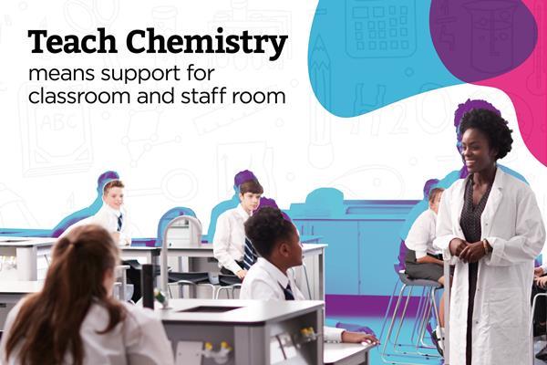
Register for Teach Chemistry for free, unlimited access
Registration is open to all teachers and technicians at secondary schools, colleges and teacher training institutions in the UK and Ireland.
Get all this, plus much more:
- unlimited access to resources, core practical videos and Education in Chemistry articles
- teacher well-being toolkit, personal development resources and online assessments
- applications for funding to support your lessons
Already a Teach Chemistry member? Sign in now.
Not eligible for Teach Chemistry? Sign up for a personal account instead, or you can also access all our resources with Royal Society of Chemistry membership.