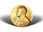
Venkatraman Ramakrishnan, Thomas Steitz, and Ada Yonath have won the 2009 Nobel Prize for chemistry for mapping the ribosome at the atomic level

Venkatraman Ramakrishnan of the MRC Laboratory in Cambridge, UK, Thomas Steitz of Yale University, US, and Ada Yonath of the Weizmann Institute of Science in Rehovot, Israel, have won the 2009 Nobel Prize for chemistry for their pioneering research on the structure and function of the ribosome - the protein-producing particle inside all cells.
The Nobel committee has described the 2009 chemistry prize as representing a third part in a trilogy that takes us from Darwin's theory of natural selection and evolution to an understanding of life at the molecular level. It was Rosalind Franklin's x-ray images of DNA that led to the 1962 prize for James Watson, Francis Crick, and Maurice Wilkins and their atomic model of the double-stranded DNA molecule, and in the 2006, it was x-rays again that showed Roger D. Kornberg how information is copied from DNA to the messenger RNA (mRNA) molecule. The ribosome structure completes the trilogy. The recipients of this year's prize each receive an equal share of the 10 million Swedish krona (ca £ 860,000) prize money for using x-ray crystallography to map the ribosome at the atomic level.
Making proteins
Ribosomes make proteins by stringing together individual amino acids. To know which order to link the amino acids, the ribosomes use the genetic code contained in DNA. However, ribosomes cannot read the DNA double helix directly, the cell must first 'unzip' the DNA and transcribe it into the single-stranded messenger molecule, mRNA.
The ribosomes bind to mRNA and use it as a template for the correct sequence of amino acids in a particular protein. Each set of three bases codes for a particular amino acid in the mRNA. The amino acids are attached to another molecule of RNA, transfer RNA (tRNA), which enters one part of the ribosome and binds to the mRNA sequence. Another part of the ribosome does the actual chemical joining of the amino acids. This whole process lies at the heart of protein production, and reproduction processes.
There are myriad proteins in the body with their own function and form. They include the oxygen-transporting haemoglobin, the immune system's antibodies, biochemical regulators such as hormones, the collagen of the skin, connective tissues and muscles, and enzymes that speed up biochemical reactions, digest our food and detoxify poisons in our livers.
Ramakrishnan, Steitz and Yonath spent many years working to get a clear picture of what the ribosome looks like at the atomic level by using X-ray crystallography.
Work begins
Ada Yonath led the way. At the end of the 1970s, she decided to try to produce x-ray structures of the ribosome. She was faced with an immediate problem. She had to obtain good enough crystals of the protein for analysis, which is almost an impossible task for such large, complicated molecules.
Yonath turned to microbes that can withstand extreme conditions of salt water, or of high temperatures up to 75°C, or of the temperature of liquid nitrogen. She reasoned that to survive such harsh conditions, the ribosomes in these microbes would also have to be resilient and so should form stable crystals.
In 1980, Yonath published a rough structure for a ribosome. With that in hand, other researchers recognised what might be possible, and Venkatraman Ramakrishnan and Thomas Steitz joined the race to publish a high-quality ribosome structure. All three published their work almost simultaneously in 2000. First, Steitz published the structure of the 50S ribosome subunit from Haloarcula marismortui. Soon after, Yonath revealed the structure of the 30S subunit from a heat-loving microbe, Thermus thermophilus. Ramakrishnan then published an even more detailed structure. By May 2001, other researchers had begun to build on these structures and had reconstructed the T. thermophilus 70S particle at an incredible resolution (5.5Å).
As the body of knowledge on how to crystallise and analyse ribosomes increased, those from other species also became accessible to X-ray crystallography. In November 2005, the structure of ribosome from the gut bacterium Escherichia coli was published and just two weeks later another team published the structure of the E. coli protein-conducting channel bound to a translating ribosome. This structure showed the ribosome during the transfer of a newly synthesised protein strand into the protein-conducting channel.
The first structures of ribosomes complexed with tRNA and mRNA molecules were revealed in 2006. Two independent research teams had used X-ray crystallography to solve the structures and to show the functional interactions and rearrangements at high resolution for the 70S ribosome-tRNA complex. Yet other complexes and active arrangements have been revealed since, all building on the initial findings of this year's Nobel chemists.
Understanding the innermost workings of the ribosome is not only important for a scientific understanding of life but also represents a practical target. Indeed, the bacterial ribosome is the target of half of all known antibiotics and remains a structure of major therapeutic importance. This year's chemistry laureates have all generated three dimensional models showing how different antibiotics bind to the ribosome and so provided drug discovery scientists with yet more information on targeting bacterial ribosomes.
David Bradley is a freelance science writer based in Cambridge


No comments yet