New method for printing artificial bone using ink made from living bone could aid reconstructive surgery

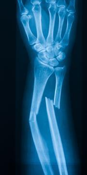
Bone takes a long time to grow and repair, so treating serious damage or carrying out reconstructive procedures can be a slow and painstaking process. However, Jake Barralet of the faculty of dentistry at McGill University in Montréal, Québec and Uwe Gbureck of the department for functional materials in medicine and dentistry at the University of Würzburg, Bavaria and their colleagues have developed a method for 'printing' artificial bone from the same chemical components as living bone using a modified ink-jet printer.1 Their approach allows them to include the biomolecules needed to trigger blood-vessel growth in the artificial material to allow it to be incorporated into living bone after it has been grafted. The printer can be used to print directly layer upon layer of artificial bone for quick-fix grafts used in reconstructive surgery.
The team used the calcium phosphate minerals brushite (CaHPO4·2H2O) and hydroxyapatite (Ca5(PO4)3(OH)) as the 'ink' in their printer. By printing one layer on top of another they can build up a highly porous material at room temperature, something that was not previously possible without strong heating. The result is a three dimensional bioceramic that closely resembles bone itself.
The addition of chemicals found in the body to stimulate blood vessel growth - vascular endothelial growth factor (VEGF) and copper sulfate - makes their bioceramic even more like bone. Tissue growth, explain the researchers, is guided by a whole range of cellular signalling molecules that ebb and flow over time, switching on and off yet more molecules that trigger growth, and crucially, growth of blood vessels into a tissue. These additives help control this process and allow blood vessels to grow into the bioceramic to help incorporate the material into the graft site completely.
Regenerative medicine is a growing field, explains Barralet, and researchers are developing the various techniques that will allow them to construct tissues, including bone, muscle, and even whole organs, outside the human body, for subsequent implantation. The biochemistry of tissue repair and integration of such engineered tissues does, however, complicate what would otherwise be a straightforward process of simply grafting the newly grown tissue onto a damaged site.
'Our low-temperature direct approach offers several practical advantages and may find application in bone grafting', the researchers say. The team has so far tested blood vessel growth into the implant materials made with and without VEGF. They found that blood vessels can grow only one or two millimetres into the pores of the artificial bone without the VEGF additive. In contrast, the artificial bone made with added VEGF promotes blood vessel growth throughout its network of pores. Such a demonstration bodes well for the further development of bespoke printable bone grafts. The researchers added that this reconstructive process could be much more effective and less risky than removing sections of bone from elsewhere in a person's body and grafting those onto an injured site.
References
- J. Barralet et al, Adv. Mater.,2007, in press.


No comments yet