


Molecular structure, especially biomolecule structure, is tricky to visualise. New resources help students see in 3D and contextualise their knowledge at the chemistry–biology interface
Molecular structure is an inherently 3D subject. This is true for small molecule chemistry, but even more so when it comes to proteins and DNA – the larger molecules of biology. Students often approach biochemistry with fear and trepidation, especially protein structure.
Proteins are large and complex molecules that refuse to be flattened. Even the simplest structure element of a protein, an alpha helix or a beta sheet, comes alive when taken out of the 2D textbook world and explored in the 3D world of computer graphics.
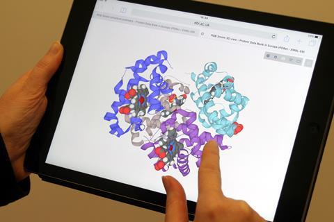
The Protein Data Bank in Europe (PDBe) contains tens of thousands of freely available real-life examples of protein structures. They are primarily used by academics and industry across the world for biological, biochemical and medical research. But, now there’s a set of resources for you.
Curriculum relevant
The resources support teaching and learning protein structure, function, and application for post-16 biology and chemistry. We’ve designed them to be flexible for either student self-study or teacher support. Resources include theory sheets, formative worksheets, quiz sheets and revision maps. There’s also a series of short videos that demonstrate some of the key learning points using animated protein structures.
The resources cover the major topics found at this level in British and Irish chemistry and biology specifications, for example:
- amino acids, peptides and protein structure;
- optical isomers, stereospecificity and computational chemistry;
- enzymes, catalysts and active sites;
- receptors, drugs and inhibitors;
- rates of enzyme reactions;
- haemoglobin and insulin;
- DNA structure and bonding, and cisplatin;
- gene editing, CRISPR and bioethics.
Ideas for the classroom
There are many ways to integrate the resources into lessons. Consider assigning them as homework or for flipped learning exercises, with students learning the theory independently before exploring structures and worksheets within the classroom. They are also well suited for revision, as summative theory sheets, revision quizzes, and revision maps all consolidate learning.
Real data
The worksheet resources make use of structures found at PDBe. Students and teachers can explore and interact with real-world scientific research data, while developing their understanding. Barriers to visualising 3D structures can be large. We have simplified the process so that students and teachers can focus on the biochemical principles.
Proteins available at PDBe include those that might be in exam specifications, such as haemoglobin or insulin. You can also see what all those proteins that appear in the news really look like, for instance the Zika virus, proteins in the CRISPR system and protein (and DNA) in a complex with anti-cancer drugs like cisplatin.
Each structure has its own webpage where you can explore it in 3D. PDBe’s online viewer, LiteMol, allows anyone to investigate molecules within a web browser, and works on any device, including tablets and smartphones. Further guides on how to access and use these are on our website.
You might also like resources developed with the Cambridge Crystallographic Data Centre for topics that require visualising 3D structures of molecules and complexes.
Matthew Conroy is a structure curator and former outreach lead at the Protein Data Bank in Europe. Peter Hoare is a STEM outreach officer at Newcastle University. Adam Stubbs is a chemistry undergraduate at Newcastle University: he has collated and developed PDBe resources, and produced a supporting series of videos.


No comments yet