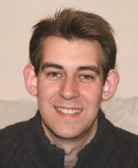
Chris has been working in the Nottingham Nanotechnology and Nanoscience Centre (NNNC) at the University of Nottingham since November 2009. He talks to David Sait about his work

Chris is an expert in cryogenic electron microscopy. Using a tool called a focussed ion beam scanning electron microscope (FIB-SEM), he is able to examine very small objects which are usually invisible to the naked eye.
Whereas a normal microscope relies on light reflected from an object to make it visible, an SEM bounces a stream of electrons onto the item, and an image is built from the electrons that bounce back. Typical objects that Chris can observe are only a few microns in size (one micron is 0.001 mm). The process also generates x-rays. These allow Chris to identify what the material is made of so he can find out chemical and structural information.
The scientific process
The NNNC is an interdisciplinary collaborative centre at the University and it supports research across a variety of disciplines including physics, chemistry, pharmacy and engineering. As a result, a lot of the research that Chris is involved with is outside his own particular area of expertise. He often needs to get to grips with an unfamiliar topic, so he spends time reading around the subject so he can understand the samples he has and how he might characterise them.
During a typical day, Chris could start by meeting PhD students, postdoctoral researchers or other academic staff to discuss the samples they need to image and the information they require. He needs to understand the nature of the sample and what the researcher’s requirements are so he can decide on the most appropriate methods to obtain the images.
There is no such thing as a standard sample or method for analysing it. Chris uses a scientific, iterative, process to obtain images and make sense of the information.
The microscopes operate under a very high vacuum, so biological samples and those from food and plant science are often cryogenically frozen in liquid nitrogen, preventing the water inside them from evaporating. This preserves the structure and shape of the object under investigation. Chris can cut into the samples using a focussed ion beam inside the microscope and take images of the internal structures that have been revealed.
Recently Chris has worked with researchers from the food sciences department. They are looking at new, greener methods of extracting oil from seeds. They asked Chris to try and obtain images of the inside of the seed, so they can identify where the oil particles are stored and what they look like.
This information will allow the researchers to improve the technologies they are working on for isolating the oil. This new process will be much more environmentally friendly as it won’t rely on organic solvents for extracting the oil.
Seeing is believing
There are many methods for measuring the size of particles, however Chris says that actually being able to see them means that we can understand them better. He also says that his favourite part of his job is knowing that he is one of the very few people in the world who can see matter on such a small scale.
Pathway to success
2009–present, research officer at the Nottingham Nanotechnology and Nanoscience centre
2008–2009, lecturer in chemistry at City College Coventry
2005–2008, postdoctoral researcher in biosciences at the University of Warwick
2002–2005, PhD in polymer chemistry at the University of Warwick
1998–2002, MChem with german at the University of Hull
1996–1998, Chemistry, geology, german and french A-levels, and mathematics AS-level, at Seevic Sixth Form College in Essex
This article was originally published in InfoChem


No comments yet